The human vertebral column contains 33 vertebrae, classified into five types from head to hip: 7 cervical (neck), 12 thoracic (chest/rib-bearing), 5 lumbar (lower back), 5 sacral (fused into the sacrum), and 4 coccygeal (the tailbone, also typically fused). Adults have 24 separate vertebrae, because the sacral and coccygeal groups fuse into solid bones in the womb.
The skeleton is what supports and anchors the soft tissue of our body, such as muscles and skin. It gives mobility and a definite shape, as well as protecting us from environmental stressors. For example, the skull protects the brain, while the ribs protect the lungs and the heart.
The Skeletal System
It should be obvious that the skeleton is one of the most important parts of our body. The skeleton is made up of bones – tissue composed of structural proteins, such as collagen and minerals, primarily calcium, as well as various bone cells and blood vessels.
There are 206 bones that form our skeleton. These 206 bones can be divided into the axial and appendicular skeleton. The axial skeleton is comprised of the bones that make up our face, neck and backbone (the vertebrae). You can remember this by recollecting that the word axial means a line (imaginary or real) at the center of an object. All bones in the axial skeleton are those which the central axis of the body could pass through.
The appendicular skeleton is formed of all the bones other than the axial bones, such as the bones that form the arms and legs. The word “appendicular” means “limbs”.
In this article, we will look at a particular set of bones within the axial skeleton – the vertebral column. This is the column through which our spinal cord passes, an essential aspect of our existence!
There are five different types of vertebrae:
- Cervical vertebrae (neck, 7 bones)
- Thoracic vertebrae (chest, 12 bones)
- Lumbar vertebrae (lower back, 5 bones)
- Sacral vertebrae (5 bones, fused into the sacrum)
- Coccygeal vertebrae (4 bones, fused into the coccyx, the tailbone)
The sacral and coccygeal vertebrae are sometimes lumped together as the sacrococcygeal region, which is the approach we’ll take below.
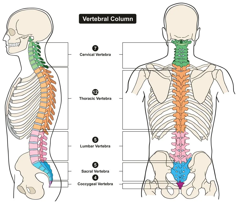
Vertebral Column
Before we delve into the different types of vertebral bones, it’s important to understand the structure and features of all vertebral bones.
The vertebral column picks up from where the skull ends and goes right down to the lower back. It is composed of 33 individual vertebrae (developmentally; in adults, the lower 9 fuse into the sacrum and coccyx, leaving 24 free vertebrae), and the entire column is divided into 5 sections – cervical, thoracic, lumbar, sacrum and coccyx – in descending order of location. Each section has a different number of vertebrae, interspersed with intervertebral discs. These discs allow for movement, act as shock absorbers, form fibrocartilaginous joints, etc. They are an important part of the vertebral column.
The typical structure of a vertebra is illustrated below:
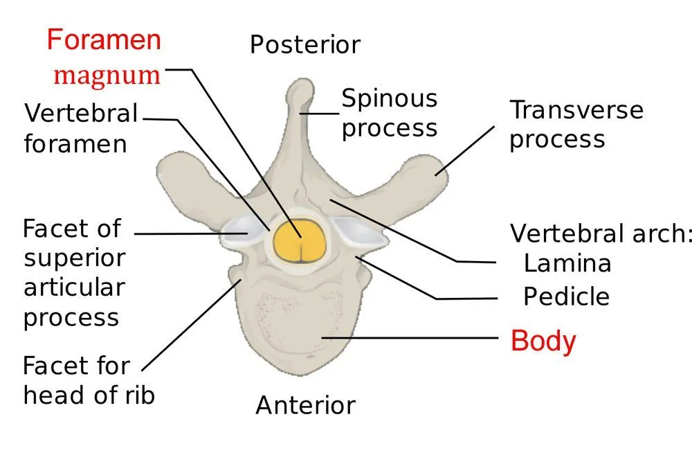
There is a part of the body, the vertebral foramen, where the spinal cord, the process that handles nerves from the brain, passes through. Several other processes exist in the vertebral bone, such as the spinous process, seen in the picture above. These processes are elongated, bony protrusions that help in association and articulation with muscles and other bones.
Cervical Vertebrae
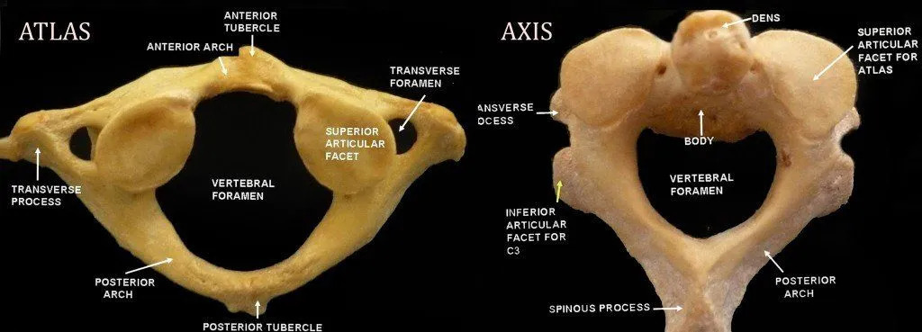
These are the bones that form the neck, making up the first section of the vertebral column. They comprise 7 bones – C1 to C7 (this is the naming convention for vertebral bones).
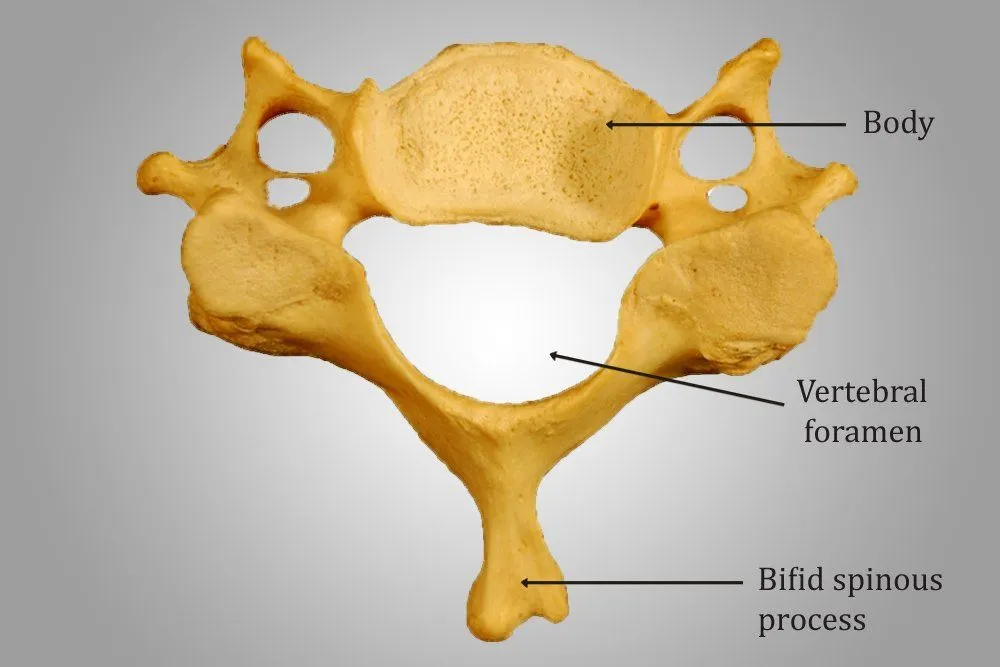
They generally have a smaller body and larger vertebral foramen, and are the smallest vertebrae in the spine. Their spinous process is bifid, meaning that the spinous processes are divided into two segments. In the cervical vertebrae, the two segments are unequal.
While the structure of C3 to C6 is similar, C1, C2 and C7 are slightly different, as they are modified for specific functions.
C1 is called the atlas. It has this name because it bears the skull, considered the “globe” of the body. The atlas lacks a body and bears a ring-like structure. It has a wide vertebral foramen to accommodate the thick part of the spinal cord.
Below this is the C2, called the axis. This acts as a pivot upon which the atlas rotates. The most notable part of this bone is the odontoid process, which rises perpendicularly. It is this part that breaks during a ‘judicial hanging’, hitting the medulla and causing death.
If you touch the nape of your neck, a bony protrusion can be felt. This is the C7, also called the vertebra prominens. The protrusion is the spinous process, which is not bifid, of C7.
Thoracic Vertebrae
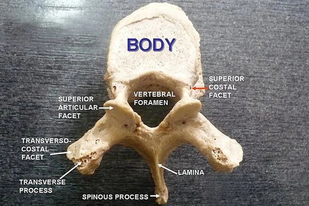
There are 12 thoracic vertebral bones, referred to as T1 – T12.
When you think of the thoracic region, the first thing that comes to mind is the rib cage. The rib cage lies in the thoracic region of the body, and the thoracic vertebrae have a slight modification in order to articulate with the ribs. This modification comes in the forms of demifacets. Facets are basically flattened surfaces of a bone. A demifacet is half of a facet, and is meant for articulation with the ribs and coastal cartilage. The vertebrae have both superior and inferior demifacets. Each rib articulates with the corresponding vertebra and the vertebra above it.
Lumbar Vertebrae
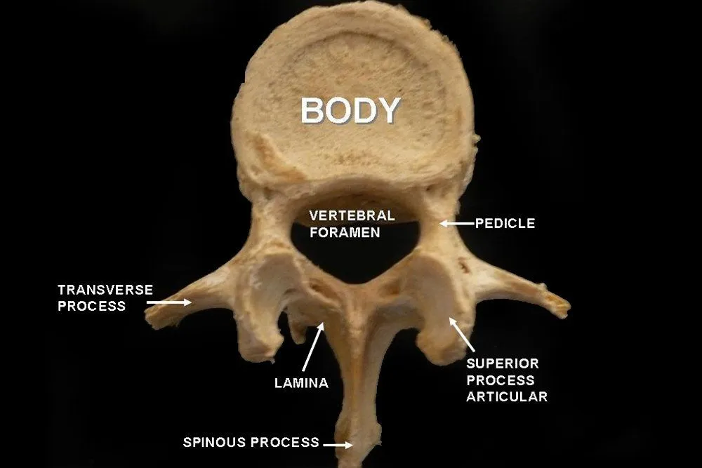
There are five lumbar vertebrae, designated as L1 – L5.
These are the largest vertebrae and have huge bodies. They bear the weight of the body and have a great capacity for movement. The orientation of their process is parasagittal (adjacent to the sagittal or longitudinal plane), which is supposedly responsible for their bending capacity. Spondylosis is most common in this region of the vertebral column.
Sacrococcygeal Vertebrae
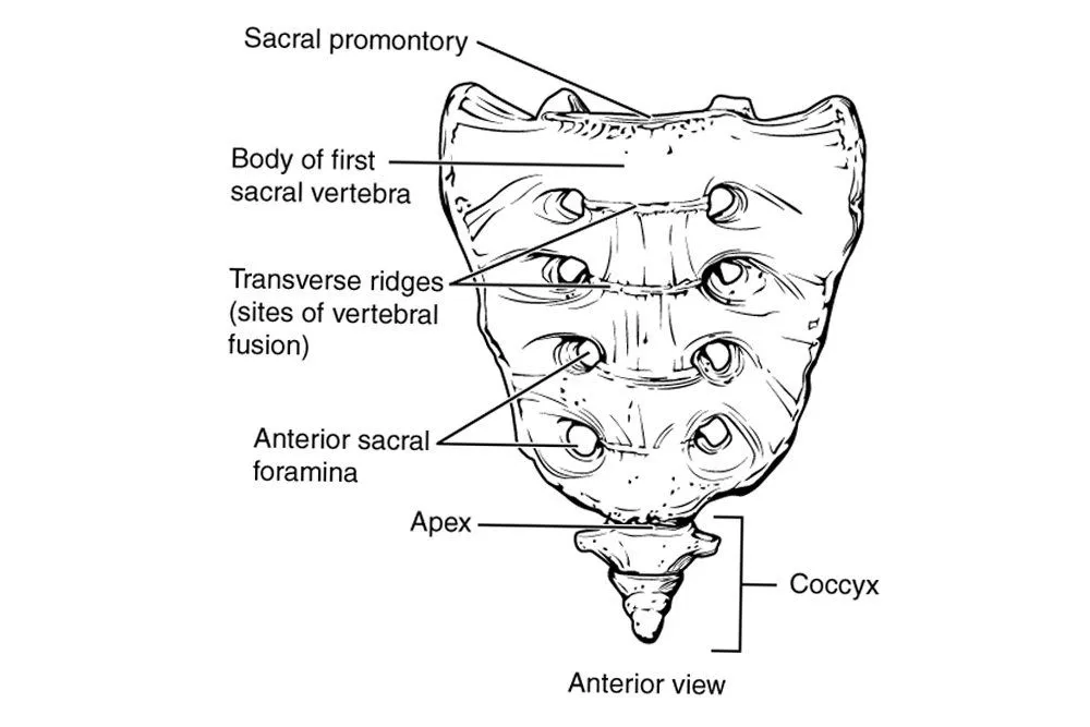
More often than not, the sacrum and coccyx are studied together as the sacrococcygeal region. The sacrum consists of 5 bones that fuse together to form a single bone, while the coccyx consists of 3-5 bones that typically do the same. In females, the sacrum is short and wide, whereas in males, it is long and narrow. The coccyx is the vestigial remnant of the tail.
How To Tell Cervical, Thoracic And Lumbar Vertebrae Apart
Once you have seen a few vertebrae, a natural question follows: if someone handed you a single loose bone from the spine, how would you know whether it came from the neck, the chest or the lower back? Anatomists rely on a short checklist of features, because each region carries a giveaway that the others do not share.
The clearest clue belongs to the cervical vertebrae. They are the only vertebrae with a transverse foramen, a small hole in each of the side (transverse) processes through which the vertebral arteries travel up to the brain. Cervical vertebrae are also the smallest, with a modest body and a triangular vertebral foramen, and their spinous process is usually bifid (split into two) from C3 to C6.
The thoracic vertebrae give themselves away through the ribs. Each one carries costal facets (small demifacets on the body plus facets on the transverse processes) where the ribs attach, a feature no other region has. Their spinous processes are long and angle sharply downward, overlapping like roof tiles, and the vertebral foramen is more circular.
The lumbar vertebrae are recognized mainly by size and by what they lack. They have the largest, kidney-shaped bodies in the column, built to carry the weight of the upper body, and they have no transverse foramina, no costal facets and no bifid spinous process. Instead their spinous processes are short, thick and point almost straight backward.

| Feature | Cervical (C1–C7) | Thoracic (T1–T12) | Lumbar (L1–L5) |
|---|---|---|---|
| Number | 7 | 12 | 5 |
| Body size | Smallest | Medium | Largest, kidney-shaped |
| Transverse foramen | Yes (unique) | No | No |
| Rib (costal) facets | No | Yes (unique) | No |
| Spinous process | Short, often bifid | Long, angled downward | Short, thick, horizontal |
| Vertebral foramen | Triangular | Circular | Triangular |
What Type Of Bone Is A Vertebra?
If you have ever been asked what kind of bone a vertebra is, the answer is an irregular bone. Anatomists sort the bones of the body by shape into a few broad groups: long bones (such as the femur in the thigh), short bones (such as the cube-shaped bones of the wrist), flat bones (such as the ribs and many skull bones) and irregular bones. Some schemes add a fifth group, the sesamoid bones, which form inside tendons, the kneecap being the best-known example.
Irregular bones are the catch-all category for bones whose complex shape does not fit neatly into the others. A vertebra is a textbook example: it combines a rounded body, a ring of bone around the spinal cord and several bony projections into one intricate unit. Like other irregular bones, a vertebra is built mostly from spongy (cancellous) bone wrapped in a thin outer shell of compact bone, a structure that keeps it relatively light while still resisting the compressive loads that run down the spine.

Atlas Vs Axis: The First Two Cervical Vertebrae
The top two cervical vertebrae, the atlas (C1) and the axis (C2), are so specialized that they look almost nothing like a typical vertebra, and together they are the reason your head can move so freely.
The atlas is essentially a bony ring. It has no vertebral body and no spinous process, just an anterior and a posterior arch. Its upper surface cradles the two rounded occipital condyles at the base of the skull, forming the atlanto-occipital joint. This is the joint that lets you nod your head to say “yes”.
The axis sits directly below and solves a different problem. Projecting upward from its body is a peg of bone called the dens, or odontoid process. The ring of the atlas pivots around this peg at the atlanto-axial joint, which is the movement you make when you shake your head to say “no”. In short, the atlas carries the skull while the axis provides the pivot it turns on.
| Feature | Atlas (C1) | Axis (C2) |
|---|---|---|
| Vertebral body | Absent | Present |
| Distinctive part | Ring-shaped arches | Dens (odontoid process) |
| Main joint | Atlanto-occipital | Atlanto-axial |
| Movement allowed | Nodding (“yes”) | Rotation (“no”) |
Damage to the vertebral column can become extremely dangerous, as it houses the spinal cord. Therefore, one must be careful of even tiny things that affect your spine, such as your position while sitting, lifting weights, etc. Incorrect posture or strain while exercising might put pressure on certain nerves within the spinal cord, not only causing pain, but even leading to far-reaching and disastrous complications.
Life would not be the same (in fact, it would be impossible) without a protected spinal cord working to send signals back and forth from the body and the brain. In other words, do whatever you can to keep it safe!