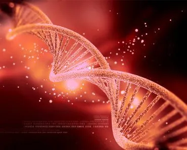
DNA itself has no color. It is colorless and transparent in solution. It only looks white when large amounts are precipitated out of a cell, where the tangled strands scatter light and where co-precipitated impurities, such as proteins, add to the white appearance.
If you were a geeky kid, you might have tried to extract DNA at home, or if not, you can certainly give it a try now. The end result of this quirky experiment would be a white gooey microscopic lump. It seems like an anticlimactic description for the extraordinary genetic material essential for all life. One might expect all sorts of funky colors from this biological essence, but sadly, that’s just not the case. Let’s look into the science behind that.
What Is The Color Of DNA?
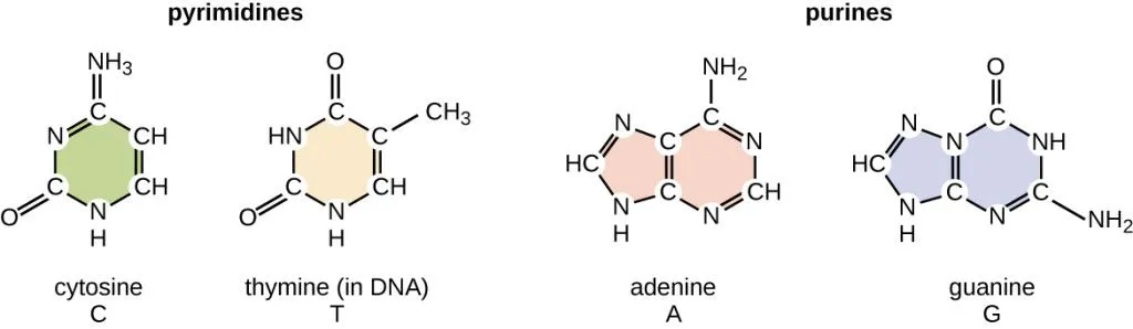
DNA is just a molecule. It’s made of a long chain of building blocks called nucleotides, which are themselves composed of smaller molecules, including sugars, phosphates, and nitrogenous bases. None of these compositional molecules are particularly colorful on their own, so it makes sense that a combination of them wouldn’t be colorful either.
Color is the result of which wavelengths of light an object absorbs and reflects back. Plants are green because they absorb red and blue wavelengths of light and reflect green and yellow back. DNA neither absorbs nor reflects visible light. This is why DNA is translucent to transparent within a cell or when dissolved in a solution.
DNA does absorb UV rays, but these unfortunately damage the DNA; absorbing UV rays causes the DNA to chemically break.
Why Does It Appear White?
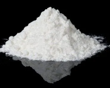
However, when we precipitate and isolate DNA out of the cell, it appears white.
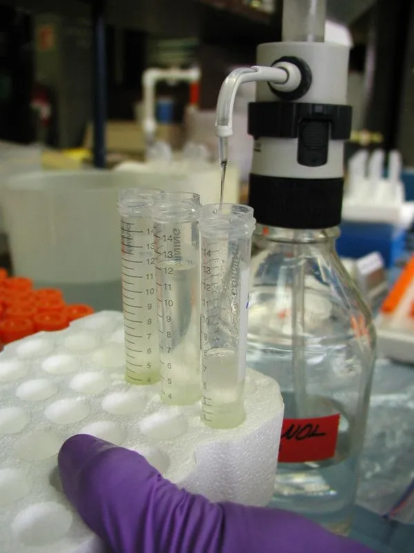
To precipitate DNA out of the water, it is mixed with cold alcohol (usually ethanol or isopropanol) along with a pinch of salt. In water, every DNA molecule is wrapped in a shell of water molecules, and its negatively charged phosphate backbone is kept apart by that water. Alcohol strips away this watery shell, and the salt provides positive ions (such as Na+) that neutralize the phosphate charges. With nothing left to keep them dissolved or apart, the DNA strands clump together and crash out of solution. This alcohol precipitation is one of the most common ways to isolate DNA.
When enough DNA is present in the solution, the precipitate forms a stringy white mass that can even be seen with the naked eye. This happens because the DNA strands become intertwined into a physical clump large enough to scatter visible light, which is what makes it appear white.
So the white color comes down to two things. First, the DNA in a precipitate is far more concentrated than it is in solution, so the tangled mass scatters light instead of letting it pass straight through. Second, a home or lab DNA extraction never pulls out perfectly pure DNA. Proteins, sugars, and other cellular leftovers come crashing out along with it, and much of the white you see actually comes from this co-precipitated gunk rather than from the DNA itself.
Interestingly, the choice of alcohol can affect how the DNA precipitate looks (both ethanol and isopropanol are themselves colorless). Less isopropanol is needed to do the job, so it tends to pull the DNA down into a denser, stringy white mass, but it also drags along more co-precipitated salt. Ethanol needs a larger volume and often yields a finer, cloudier white precipitate that is cleaner.
Can You Actually See DNA Under A Microscope?
If DNA is the molecule that makes you you, you would think you could just drop a sample under a microscope and watch that famous twisted ladder spiral away. Sadly, an ordinary light microscope will not oblige. A single DNA double helix is only about 2 nanometers wide, which works out to two billionths of a meter. A light microscope, no matter how sharp its lens, cannot resolve anything much smaller than roughly 200 nanometers, a hard limit set by the wavelength of visible light itself. That makes the helix around 100 times too thin to ever show up as a spiral under the microscopes you find in a classroom or a hospital lab.

So what about that white clump from the home extraction? What you are squinting at there is not a single molecule but many millions of DNA strands tangled together, mixed in with proteins and other cellular leftovers. Even under a light microscope, that precipitate just looks like pale, colorless threads and fibers, never a tidy double helix.
Here is the surprising part: the iconic double helix was never actually seen through a microscope at all. Its shape was worked out from X-ray diffraction, most famously in “Photo 51,” an X-ray image of DNA captured by Rosalind Franklin and her student Raymond Gosling at King’s College London in 1952. The telltale X-shaped pattern in that photo was the clue that told scientists DNA had to be a helix.
Today, researchers can image single DNA molecules directly using techniques like atomic force microscopy and electron microscopy, both of which pick out detail far below the reach of visible light. An atomic force microscope traces the surface of a molecule with an ultra-fine tip, and can map a single molecule of DNA dried onto a flat surface without using any stain at all.
Is There Another Color In Which DNA Can Be Seen?
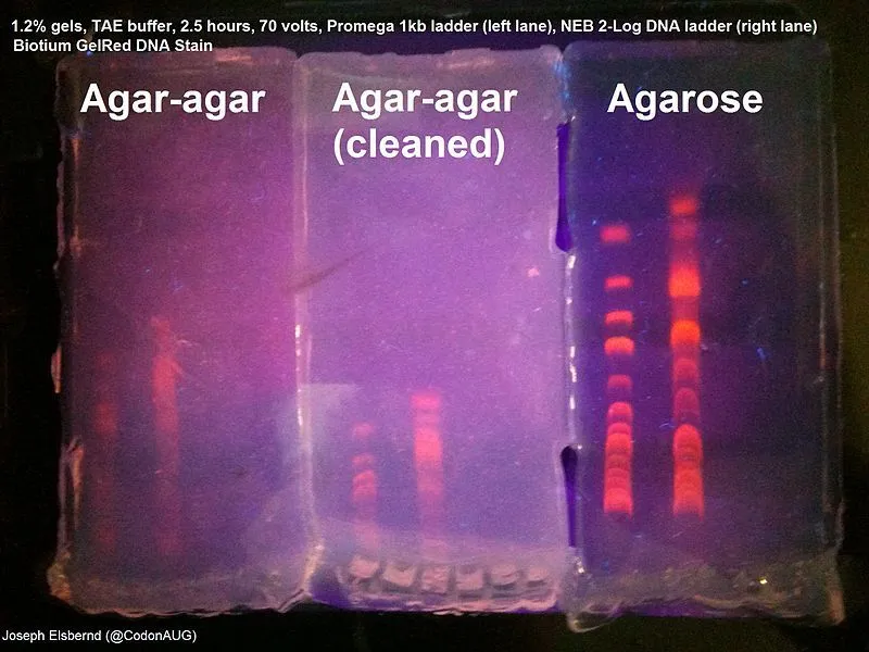
Absolutely, yes! With the help of DNA dyes, DNA can be seen in a variety of fluorescent colors. DNA dyes are colorful compounds that just love to hang out with DNA molecules. They make it easy for scientists to spot and study DNA in the lab by emitting fluorescent light. There are several types of DNA dyes, including old-school Ethidium Bromide (EtBr), which glows orange-red under UV light.
On the other hand, there is SYBR Green, which emits a cool green light and is far less toxic. GelRed is a newer dye that is similar to EB, but is a bit more well-behaved, glowing red under UV light. Hoechst dyes are like the shy kid who only hangs out in the minor groove of DNA, and glows blue under UV light.
Basically, the choice of dye depends on what the scientists are studying and what they wish to observe with the coloration. Some dyes may be more sensitive or specific to certain types of DNA, while others may be easier or safer to use. One thing is for sure: without these colorful characters, it would be much harder to study the fascinating world of DNA.
Why Do DNA Dyes Glow Under UV Light?
We just saw that dyes let us watch DNA in living color, but where does that glow actually come from? The answer is a process called fluorescence. A fluorescent molecule soaks up high-energy light, in this case invisible ultraviolet, and almost instantly throws part of that energy back out as lower-energy light our eyes can see. The dye absorbs UV and re-emits it as visible orange, red, green, or blue, depending on the dye. So when a stained gel lights up on a UV lamp, it is the dye that is glowing, not the DNA. Pure DNA, remember, neither reflects nor emits visible light of its own.

Take ethidium bromide, the old-school classic. It is a flat molecule that wedges itself, or intercalates, between the stacked rungs of the double helix. Sliding into that snug pocket does something striking: the dye fluoresces far more brightly than it does when floating free in solution. The reason comes down to water. Out in the surrounding liquid, each dye molecule is wrapped in water molecules, which are very good at quietly draining away (quenching) its glow. Tucked between the base pairs, the dye sheds that watery shell, escapes the quenching, and is finally free to shine under UV.
That is exactly why a gel shows crisp bright bands right where the DNA sits. The dye piles onto the DNA and glows hard, while the empty stretches of gel, with no DNA to bind, stay dark. It is a neat reminder that the color you see is always the dye talking, with the colorless DNA quietly underneath.
Conclusion
The ability to visualize and analyze DNA molecules has revolutionized the fields of genetics and molecular biology. It has led to incredible breakthroughs in medicine and biotechnology. By using dyes and precipitation methods to visualize DNA, scientists have been able to study the structure and function of DNA molecules, our building blocks of life. It has led to the development of new technologies, such as DNA sequencing, gene editing, and genetic engineering. The beauty and complexity of DNA are truly remarkable, and our newfound ability to see and study it has expanded our understanding of life itself.
References (click to expand)
- How To Extract DNA From Anything Living. University of Utah
- Haines, A. M., Tobe, S. S., Kobus, H. J., & Linacre, A. (2015, February 23). Properties of nucleic acid staining dyes used in gel electrophoresis. Electrophoresis. Wiley.
- Isopropanol Precipitation of DNA. QIAGEN Bench Guide
- What color is DNA? The Tech Interactive
- Pyne, A. L. B., et al. (2021). Atomic force microscopy: a tool for structural and translational DNA research. APL Bioengineering.
- Photograph 51, by Rosalind Franklin (1952). The Embryo Project Encyclopedia, Arizona State University.
- Double Helix. National Human Genome Research Institute (NHGRI).
- Olmsted, J., & Kearns, D. R. (1977). Mechanism of ethidium bromide fluorescence enhancement on binding to nucleic acids. Biochemistry.